Clinical Image & Video Gallery
When constituted, ExEm® Foam produces approximately 127,000 micro air bubbles, making the image bright and white, providing a clear view of where the foam is. When the foam is injected into the uterus and tubes, patent fallopian tubes will appear as a thin, echogenic (bright white) line, when visualized with ultrasound. If the white line does not appear, the tubes might be occluded. ExEm® Foam is the only FDA-approved contrast agent for use with Trans Vaginal Ultrasound (TVUS), which can be performed as an in-office procedure.

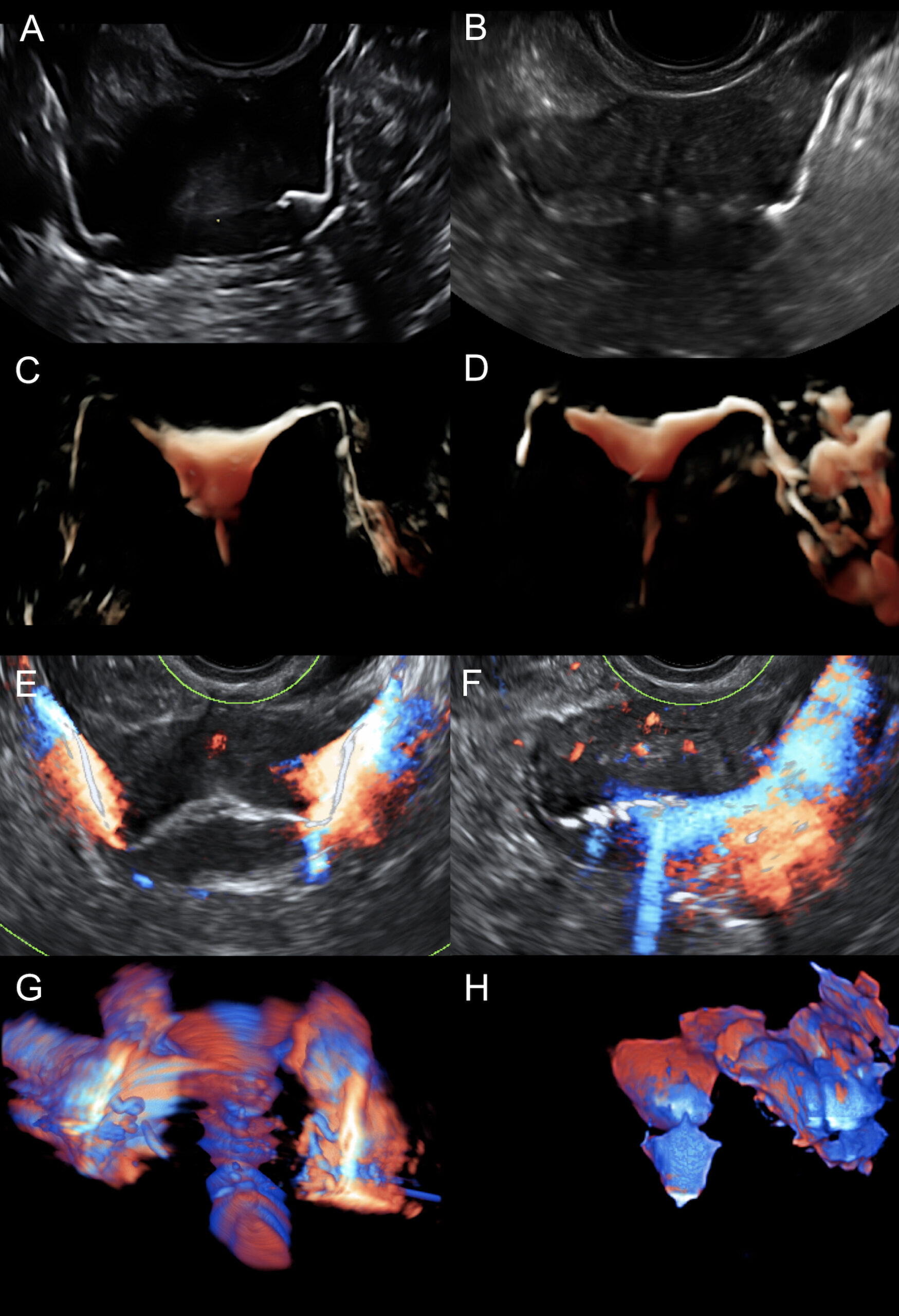
ExEm® Foam images of the uterus with the fallopian tubes in two women with known or suspected infertility. The left column shows patency in both fallopian tubes and the right column shows occlusion in left tube.
Technology used (top to bottom):
• 2D-HyFoSy (A+B)
• Offline HD-live rendered
• 3D-HyFoSy (C+D)
• 2D-HDF-HyFoSy (E+F)
• Offline color-rendered
• 3D-HDF-HyFoSy (G+H)
(Source: Ludwin I, et al. 2017)
Images (E) through (F): if the foam flow is present throughout the catheter, the uterus, and the patent tube during infusion, positive contrasting by the foam is significantly enhanced by color (left column). One fallopian tube is occluded in the right column: in (B) and (D), it is partially contrasted by foam, and in (F) and (H), it is not contrasted by color.
(Source: Ludwin I, et al. 2017)


